Thalia Rehab Project: Examination of the Laminitic Hoof
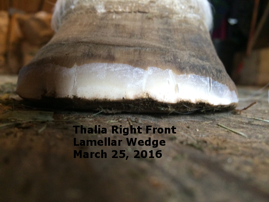
This is the 3rd trim since Thalia joined my barn around the first of the year. An improved connection of her hoof capsule to the coffin bone (P3), which you can see in the “Right Front” photo, has grown down from the coronary band enough for me to feel comfortable with REALLY backing her toe up. I used my nippers laying flat on the barn floor to clip away dead hoof. This is the rough cut before rasping smooth. Saves me lots of rasping! What you see in the picture is all dead hoof, called the Lamellar Wedge . The lamellar wedge is what forms in between the live structures of the inner hoof and the hoof capsule. In other words the “white line”. That thin yellow/whitish line between the hoof wall and the sole. This is what happens to the white line when the coffin bone loses its connection to the hoof capsule. The white line in the area where separation has occurred stretches. And the more it stretches the more severe the rotation. The more severe the rotation the larger the lamellar wedge will be.
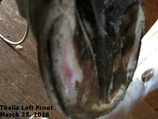
The “Left Front” picture shows how Thalia’s coffin bones have become “sinkers”. “Sinkers” are coffin bones which not only rotate out of the horizontal position toward a vertical position but also lose connection to the hoof capsule so far up toward the coronary band that P3″sinks” toward the ground. In these cases a convex instead of a flat or concave sole develops.
Notice how the left sole bulges, and how her frog has an upward curve in its shape toward the tip of the frog. This is how I know the coffin bone has not only rotated but equally as bad, dropped toward the ground away from P2. This mare was in danger of sloughing off the hoof capsule. What saved her was the tremendous amount of dead sole she had accumulated. I’m leaving all that dead sole in place for now. She needs it for support of her internal structures. The red spot you see is from previous bleeding into the live, (like your skin) sole trapped under the dead sole. The position of that red spot is directly over a section of the outer edge of the coffin bone. I’ve used pictures of both the left and the right front. They are equally poor mechanically.
On the upside, and another saving grace for her, she has very healthy frogs and bulbs in both front feet. This is most unusual in a laminitic horse. It says that her hoof mechanics, pre how ever many laminitis episodes she’s had, were good enough to make her comfortable about using the back part of her hoof in a healthy way. You don’t see strong fogs and bulbs like hers on horses who have had too much heel left after trims or shoeing year after year.
Good demonstration and explanation, Jeannie!