Laminits and And The Consequences: Thalia
Thalia’s hooves. Where do I begin?
I knew she’d had laminitis (active) in mid-October 2015 due to eyewitness reports. What I’ve discovered, now that she is with me, is she has been having laminitis attacks, off and on, for at least a year. And without supportive care. The coffin bones (P3) in her front hooves have descended so close to the ground that if the compacted sole on the bottom of her hooves were removed (which I’d normally do) her coffin bone would penetrate right through the live sole. What this means is that she has had attack after attack after attack of laminitis and her laminae have torn apart all the way up to just short of the coronary band. Laminae are much like velcro in the way the sensitive tissue adheres to the insensitive horn of the hoof wall. This happens over time, a slow insidious degenerative process.
How am I so sure of this? The state of her hoof tells me this. First lets look at the outer wall.
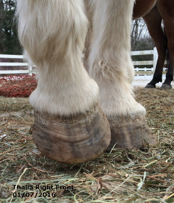
Each one of those rings represents a laminitis attack. Rings in and of themselves don’t necessarily say “laminitis”, however this clue, along with the angle of her hoof wall and the sole of her hoof scream laminitis, and not just one episode either.
Here is a look of that same hoof from the side.
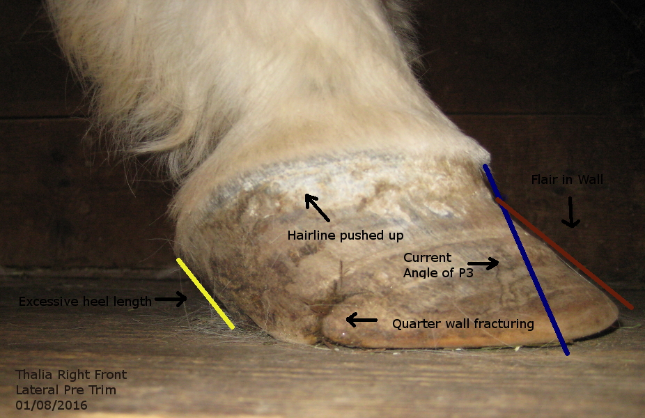
This photo shows the only tight connection of her sensitive inner hoof to the hoof capsule occurs in the top 1/2 inch of that hoof wall starting at the coronary band. The blue line shows the angle the hoof wants to grow at in order to maintain the connection of the outer wall with the inner coffin bone. The angle of growth from the coronary band tells you the angle of the coffin bone within.
The red line shows where the laminae have separated inside and resulted in what is known as flare. The triangle formed by theses two lines represents “dead” tissue inside the hoof capsule known as the lamellar wedge. The yellow line shows how long her heels are and how far forward from the back of her hoof the ground contact with the heels is. The pushed up hairline represents both the steep angle of the coffin bone and also the excessive pressure of the quarter area of the hoof on the lateral cartilages inside the hoof.
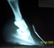
From Pete Ramey’s HoofRehab.com site this film shows what the coffin bone angle would look like inside Thalia’s hoof.
Below is what the lateral view of a front hoof should look like.
Take note of the low heels, the angle of the wall and the angle of the hairline.
Now let’s look at the underside of Thalia’s hoof. It is always a challenge to show in a picture what I can see with my eyes.
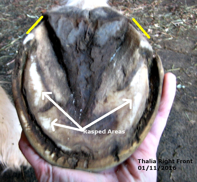
In order to try and show how the sole of her hoof is convex instead of concave I used the rasp flat across her hoof lightly to show that the only places the where the rasp touched were the outer edges of her sole, just barley the tips of her heels, and the medial (inside) wall in a couple of places. You can also see the extreme separation of hoof capsule from the inner hoof in this picture. The separation you see here is not the result of white-line disease. Meaning white-line disease is a result of rotation and laminae death not the cause of the separation in this case. The yellow lines in this picture show the length of her heels as viewed from the bottom of the hoof. On a good note she has a nice strong and un-compromised frog.
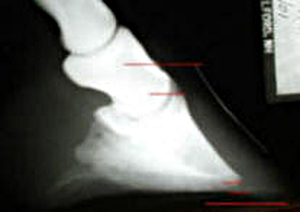
However, her coffin bone is right at ground level instead of snug up inside the hoof capsule. Hence the bulging sole. Here is another picture from the HoofRehab.com site showing what it looks like inside the hoof when this happens.
Her left front hoof looks just as compromised as her right front and her resting stance and walking movement tell me her right front is the more painful of the two front hooves.
Jeannie–have you gotten any radiographs of her feet?
No, not yet. I don’t really feel the need to. The external appearance of her hoof is telling me what I need to know to trim her. I’ll be posting the post trim pictures soon. The first trim has already taken place. I’m just slow in putting stuff up!
What do you hope to accomplish by proper care?
I will help her to grow a hoof that will establish a tight connection to the coffin bone within and will bring both the angle of the coffin bone back to a proper position and also lift it “up” deeper into the hoof capsule. I’ve done this before. A horse typically grows a hoof from the coronary band to the ground in 9-12 months. So it will take that long to achieve my goal if everything goes as planned.
I think you are treating her wrong (based upon the last picture at least)
To start, you have to take away the edges of the hoofwall up to 1-2cm from the ground – you can compare laminitis best with a loose fingernail, it hurt as hell when they touch ground with the hoofwall or bump into something.
You also have to cut away the heel as low as possible to help turning back the angle.
If you don’t have hoofshoes, you might have to tape rubber pads below the hoof in the first week.
There is an interesting video on Youtube http://www.youtube.com/watch?v=RrwRYdOiYXs – when my foundered mare got an abcess I did something similar but using a rubber pad instead of reversed iron (sliced to the size of the hoof) and I didn’t open it in front. About 4 weeks later I lost half of the sole because of pressure of the abcess – not sure if it would have helped to have opened it in front as well.
Thanks, Jeannie! Fingers crossed Thalia has a smooth course.
Hmmm, Patrick. I haven’t posted pictures here yet of how I have trimmed her. These pictures show her right front hoof as I received her. What makes you think I’d already trimmed her? By the way, she is doing splendidly with her new trim and I need to post pictures of that.